A case of multiple articular fractures
Case Study & Surgery

Sophie is an approximately 2 year old female Border Collie mix who was presented to the VSB Surgery Service with a 1.5 month history of bilateral pelvic limb lameness. The right pelvic limb lameness was historically significantly worse than the left. The lameness occurred due to an unknown trauma. Sophie was found with her injuries and no owner could be located. Sophie was initially retained by animal control before she was ultimately surrendered to a rescue organization, which then took over her care. Radiographs performed soon after her adoption demonstrated an intra-articular left femoral head fracture with coxofemoral luxation and an intra-articular right femoral trochlear fracture. Several ballistic projectiles were found scattered around the pelvis as well. There was no evidence of recent external puncture wounds, so the ballistic projectiles were presumed to have arrived during a previous traumatic event.
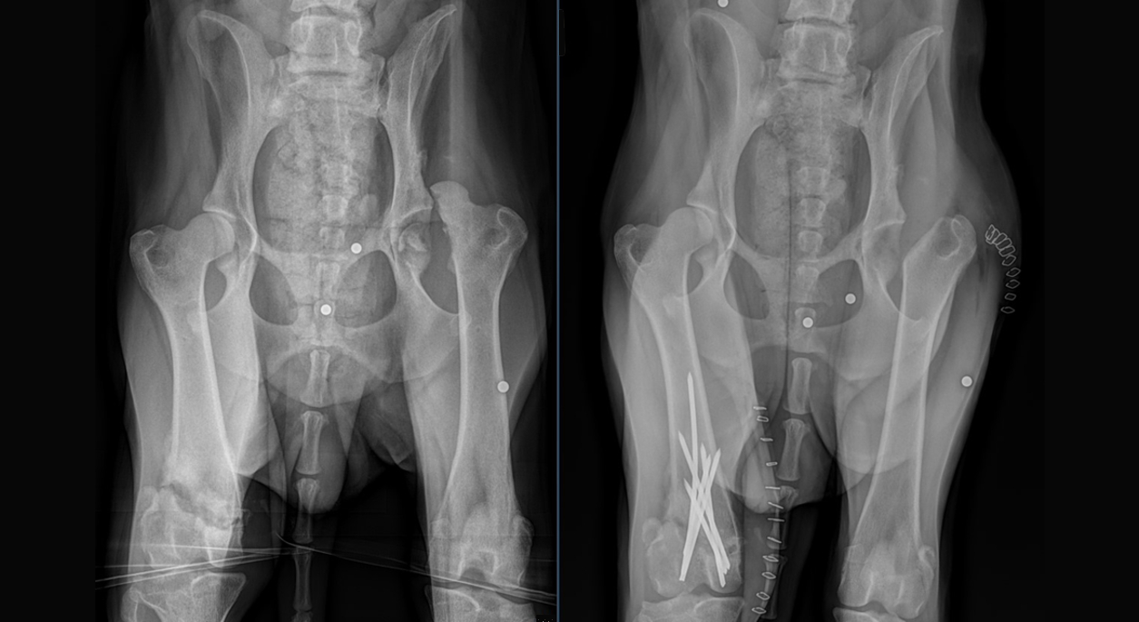
The bilateral nature of Sophie’s injuries made treatment recommendations difficult. A left femoral head and neck ostectomy was recommended for treatment of the left femoral head fracture and coxofemoral luxation. The treatment recommendation for the right femoral trochlear fracture was less clear. Amputation was considered, but fracture reduction and stabilization would likely result in a better overall function if such a procedure were possible. The relatively long time between injury and presentation raised concerns over the ability to alter the reduction and alignment of the bone fragments. Fibrosis and early bone healing could significantly inhibit attempts to alter the position of the bone fragments. After much consideration, the choice was made to proceed with left FHNO and right femoral trochlear fracture reduction and stabilization, if possible.
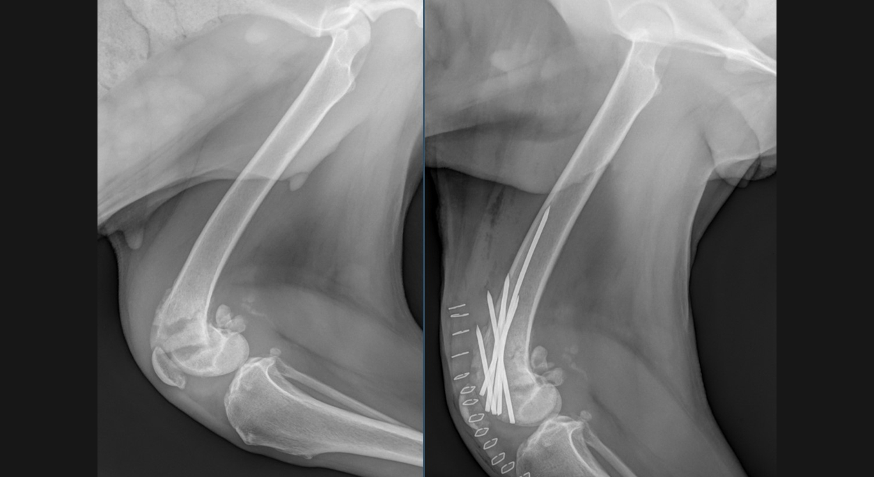
The right femoral trochlear fracture was approached and found to be moderately unstable. The fracture extended medial to lateral through the proximal femoral trochlea, resulting in articular involvement both above and below the fracture line. After severing fibrous adhesions between the bone fragments, the fracture could be reduced with significant effort. The fracture was ultimately stabilized with six IM pins placed in cross-pin fashion. Postoperative fracture reduction and alignment were considered good. The left FHNO was routine. Sophie is now on the road to recovery and will hopefully regain good function of both pelvic limbs.
