Craniodorsal Coxofemoral Luxation in a Dog
October 7, 2025 · Surgery

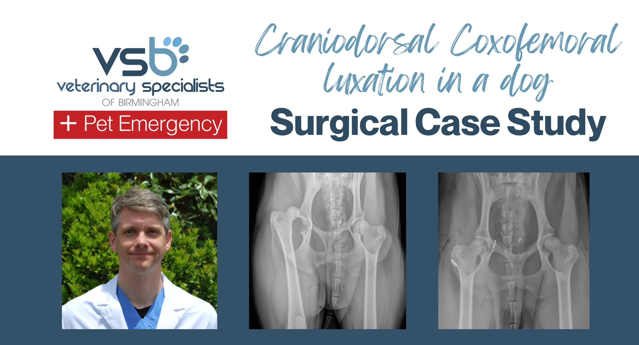
A 4-year-old male castrated Standard Poodle was presented for evaluation of acute onset right pelvic limb lameness after being hit by a car 2 days earlier. Radiographs performed by the primary veterinarian showed a craniodorsal right coxofemoral luxation. Coxofemoral congruity was good and no fractures were seen. The patient was in good overall health and no other orthopedic injuries were found.
Several treatment options exist for craniodorsal coxofemoral luxation. Closed reduction with or without Ehmer sling placement is the least invasive and generally simplest treatment option but has a >50% rate of recurrence.
Femoral head and neck ostectomy (FHNO) is a relatively simple surgical option with a good degree of tolerance in small to medium breed patients, but large breed patients and those with orthopedic co-morbidities can have a poor outcome. FHNO also has no opportunities for revision if the outcome is poor.
Open reduction and stabilization of the coxofemoral joint offers the best potential for restoration of normal joint mechanics. Numerous techniques are reported for stabilization of the coxofemoral joint following open reduction, but this author currently favors toggle rod stabilization.
Total hip replacement (THR) is an option for treatment of coxofemoral luxation but should generally be reserved for patients with poor coxofemoral congruity, recurrent luxations, or patients with significant cartilage pathology. The clients in this case chose to pursue open reduction and toggle rod stabilization.
An Arthrex TightRope implant was selected for implantation based on the patient size (25 kg). The TightRope implant is a braided high molecular weight polyethylene (UHMWPE) with a braided polyester and UHMWPE jacket. The implant has a very high tensile strength and good abrasion resistance, making it in many ways a better choice than the traditional nylon monofilament suture.
The implant was placed through a 3.5 mm tunnel in the acetabulum and a 3.2 mm tunnel in the femoral head and neck. The joint capsule was then reconstructed prior to wound closure.
